Novel ultrasound transducers/arrays for biomedical imaging
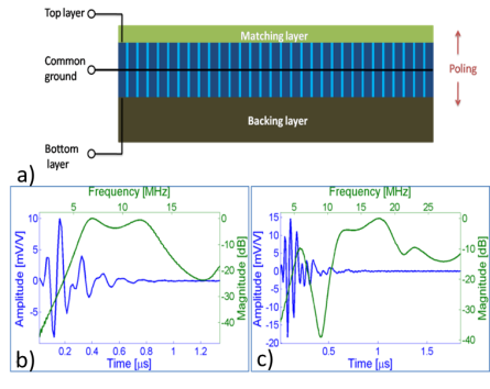
Micromachined piezo-composite transducers
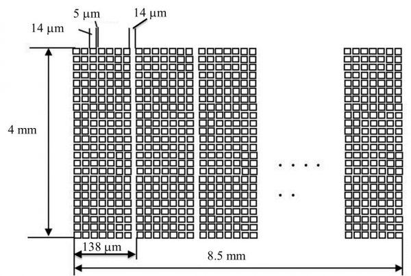
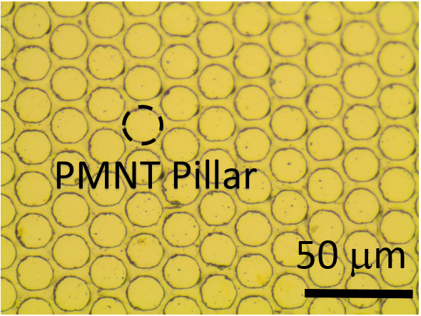
Composite piezoelectric materials have high electromechanical coupling factors, low acoustic impedance, and are relatively easy to conform. However, fabrication of high frequency composite transducers and arrays becomes difficult because of fine kerfs and fine array pitches. Recent progress on micromachined 1-3 single crystal piezoelectric composite transducer technology (PC-MUT) showed that 15-75 MHz 1-3 composites can be fabricated with excellent properties. This PC-MUT technology takes the advantage of high electromechanical coupling coefficients of advance single crystal piezoelectrics, photolithography and DRIE based fine patterning of both composite structures and array electrode interconnections. These micromachined piezo-composite transducers and arrays are promising for advanced biomedeical imaging.
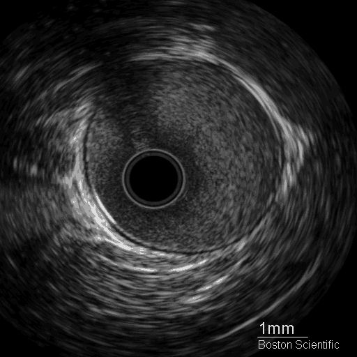
IVUS imaging using a 55 MHz micromachined 1-3 composite transducer
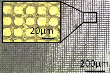
35 MHz 64-element linear array
Contrast Enhanced Superharmonic IVUS Imaging — Acoustic Angiography
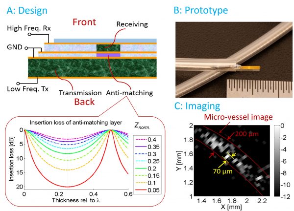
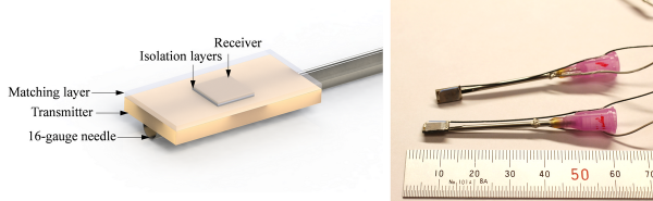
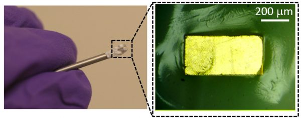
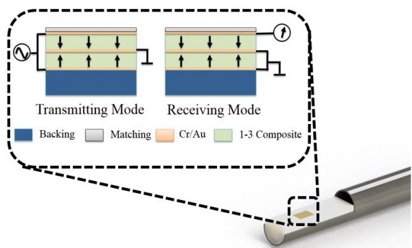
Atherosclerotic cardiovascular disease is a leading cause of death worldwide, and one which often manifests without warning. Imaging of coronary vasa vasorum may lead to better assessment of the vulnerable plaque development in diagnosis of atherosclerosis diseases. Intravascular ultrasound (IVUS) imaging transducers capable of detecting microvessels via nonlinear contrast imaging could provide valuable diagnostic information; however, such transducers are not yet available commercially. In this research, dual frequency IVUS transducers (Figure A) were developed with a low frequency (6.5 or 5 MHz) transmitter to excite ultrasound contrast agents (microbubbles) and a high frequency (30 MHz) receiver to detect the super-harmonics from microbubbles. An anti-matching layer was sandwiched between the two active elements to suppress the aliasing echo in high frequency receiving wave. The transducer prototype (Figure B) was small enough to be integrated into a commercial catheter (Boston Scientific, Natick, MA). The imaging result (Figure C) elucidated the detection of a micro-tube (mimicking vasa vasorum) with 200 µm diameter. Pulse-length of each bubble response was about 70 µm, indicating its capability of high resolution intravascular acoustic angiography.
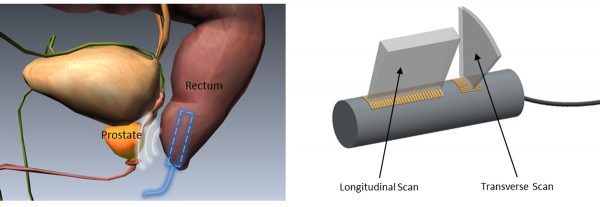
Dual frequency linear array for prostate imaging
Transrectal ultrasound (TRUS) has been considered as the standard diagnostic method for the prostate cancer assessment. In this research, a dual-layer dual-frequency and dual mode TRUS transducer for contrast-enhanced ultrasound imaging of vasa vasorum, toward acoustic angiography of prostate imaging.
The dual frequency transducer prototype with a 2 MHz 1-3 composite transmitter and a 14 MHz receiver was fabricated and characterized, followed by microbubble detection tests. The transmitter showed a peak negative pressure (PNP) of -1.5 MPa. The -6 dB pulse echo fractional bandwidth for the transmitter and receiver were 61% and 45%, respectively. The prototyped dual frequency transducer was used to successfully excite microbubbles and to detect super-harmonic responses from microbubbles. The measured harmonic signal showed a 12 dB contrast-to-noise ratio (CNR).
Dual-frequency intravascular ultrasound array
The ultrasonic contrast imaging for molecular or vasa vasorum (VV) assessment can be promising in identification of vulnerable plaques. However, conventional intravascular ultrasound (IVUS) transducers with frequency of 15 MHz – 60 MHz are not optimized for imaging with micro bubble contrast agents due to the ineffective micro bubble excitation at high frequencies. We designed and fabricated a dual-frequency catheter-based array for intravascular high resolution ultrasonic fundamental imaging and contrast enhanced acoustic angiography. A miniaturized dual-frequency array was designed as a dual layer PMN-PT structure, where the transmission part is an 8-element circular array and the receiving part is a 64-element circular array. The center frequency of low frequency transmission array and high frequency receiving array are 5-MHz and 35-MHz, respectively.
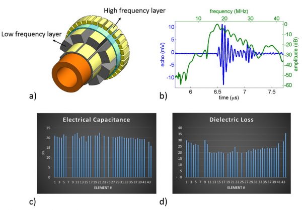
a) Sketch of dual-frequency IVUS array transducer; b) Pulse-echo waveform and frequency spectrum of receiving array; c) Electrical capacitance and; d) dielectric loss of receiving array
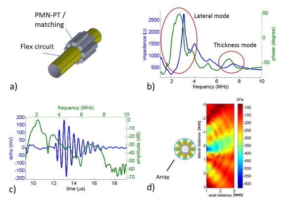
Transmitter array characterization. a) Structure of transmitter array. b) Electrical impedance measurement result. c) Pulse-echo result. d) Acoustic mapping of 5 elements, 25 Volts, 2-cycle burst excitation.
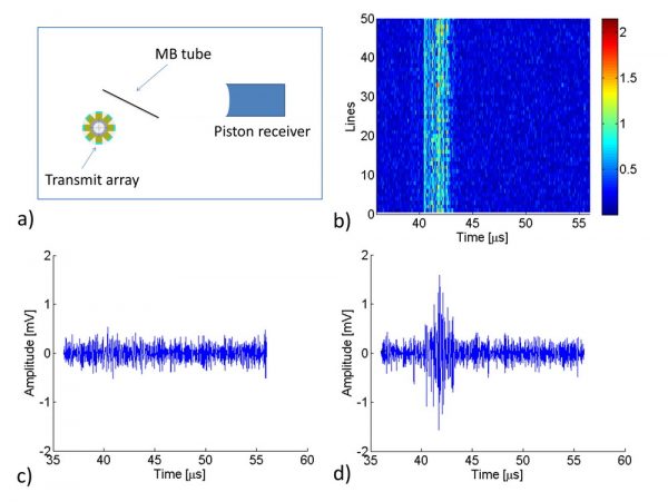
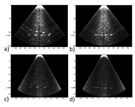
Contrast test result (5-cycle 90V excitation, 25 MHz receiver). a) Experimental setup. b) Map processed from 50 A-lines. c) An A-line without micro bubbles. d) An A-line with micro bubbles.
Bi-frequency Co-Linear Array
Ultrasound imaging with high resolution and large field of depth has been increasingly adopted in medical diagnosis, surgery guidance and treatment assessment. Conventional ultrasound works at a particular frequency, with -6 dB fractional bandwidth of ~ 70 %, limiting the resolution or field of depth in many ultrasound imaging applications. A bi-frequency co-linear array (7.5 MHz/15 MHz) covering a frequency range of 5 MHz – 20 MHz was investigated to meet the requirements of broadband ultrasound imaging.
a) Structure of 32 element array. b) KLM simulation of low frequency mode. c) KLM simulation of high frequency mode.
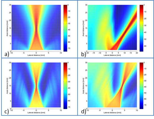
Simulation of Beamforming by using Field II program. a) The beam of low frequency was focused at 15 mm without any steering. b) The beam of low frequency was focused at 15 mm and with a 40 degree steering. c) The beam of high frequency was focused at 15 mm without any steering. d) The beam of high frequency was focused at 15 mm and with 15 degree steering.
Spatial resolution test of the 32-element co-linear array with the focal depth of 110 wavelengths (25 mm). a) The axial resolution of low frequency mode (6.93 MHz) is better than 1 mm. b) Lateral resolution of low frequency mode is better than 2 mm. c) Axial resolution of high frequency mode (15.625 MHz) is better than 0.5 mm. d) Lateral resolution of high frequency mode is better than 1 mm.
Broadband ultrasound imaging
Broadband ultrasound imaging has been widely investigated, especially for tissue harmonic imaging and contrast enhanced imaging. Recent studies aimed at exploring high frequency (> 15 MHz) broadband transducers for ophthalmology, dermatology, cardiology and small animal imaging. In general, broadband imaging possesses advantages including contrast resolution enhancement, artifacts reduction, and improved signal-to-noise ratio. Ultrasound probes with higher center frequency and broader bandwidth would provide better image resolution. This work presents the use of micromachined 1-3 composites in a dual layer, dual frequency transducer for tissue harmonic imaging applications.
References:
- Rehrig, P., X.N. Jiang, W. Hackenberger, J. Yuan and R. Romley, “Micromachined Imaging Transducer”, US Patent # 7622853, 2009.
- Jiang, X.N., W.S. Hackenberger, and K. Snook, “Micromachined Piezoelectric Ultrasound Transducer Arrays”, US Patent # 8008842, 2011.
- Jiang, X.N., K. Snook, R. Liu , X. Geng, and W. Hackenberger, “Fabrication and Characterization of High Frequency Phased Arrays for NDE Imaging”, Proc. SPIE Smart Materials and Structures and NDE, 7649-30 (2010).
- Ma, J., S. Guo, D. Wu, X. Geng, X. Jiang, “Design, fabrication, and characterization of a single-aperture 1.5-MHz/3-MHz dual-frequency HIFU transducer“, Ultrasonics, Ferroelectrics and Frequency Control, IEEE Transactions on, 60(7) (2013).
- Jian, X., S. Li, W. Huang, Y. Cui, X. Jiang, “Electromechanical response of micromachined 1-3 piezoelectric composites: Effect of etched piezo-pillar slope“, Journal of Intelligent Material Systems and Structures, 2014.
- Ma, J., K. Martin, P. Dayton, X. Jiang, “A preliminary engineering design of intravascular dual-frequency transducers for contrast enhanced acoustic angiography and molecular imaging“, Ultrasonics, Ferroelectrics and Frequency Control, IEEE Transactions on , 61(5), 870-80 (2014).
COPYRIGHT© 2012 XIAONING JIANG.
MAE, NCSU, RALEIGH, NC